CLEC10A Antibody - N-terminal region
Rabbit Polyclonal Antibody
- SPECIFICATION
- CITATIONS
- PROTOCOLS
- BACKGROUND

Application
| WB |
|---|---|
| Primary Accession | Q8IUN9 |
| Other Accession | NP_878910 |
| Reactivity | Human |
| Host | Rabbit |
| Clonality | Polyclonal |
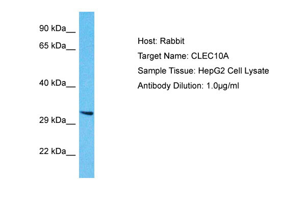
| Calculated MW | 34kDa |
| Gene ID | 10462 |
|---|---|
| Alias Symbol | CLEC10A, CLECSF13, CLECSF14, HML, |
| Other Names | C-type lectin domain family 10 member A, C-type lectin superfamily member 14, Macrophage lectin 2, CD301, CLEC10A, CLECSF13, CLECSF14, HML |
| Format | Liquid. Purified antibody supplied in 1x PBS buffer with 0.09% (w/v) sodium azide and 2% sucrose. |
| Reconstitution & Storage | Add 50 &mu, l of distilled water. Final Anti-CLEC10A antibody concentration is 1 mg/ml in PBS buffer with 2% sucrose. For longer periods of storage, store at -20°C. Avoid repeat freeze-thaw cycles. |
| Precautions | CLEC10A Antibody - N-terminal region is for research use only and not for use in diagnostic or therapeutic procedures. |
| Name | CLEC10A {ECO:0000303|PubMed:33724805} |
|---|---|
| Function | C-type lectin receptor involved in recognition of N- acetylgalactosamine (GalNAc)-terminated glycans by myeloid antigen presenting cells (APCs) (PubMed:15802303, PubMed:16998493, PubMed:17616966, PubMed:22213806, PubMed:33724805, PubMed:8598452). Binds in a Ca(2+)-dependent manner to alpha- and beta-linked GalNAc residues on glycoprotein and glycolipid antigens, including alphaGalNAc- and Galbeta1->3GalNAc-O-Ser/Thr also known as Tn and T antigens, LacdiNAc epitope GalNAcbeta1->4GlcNAc and its derivative GalNAcbeta1->4-(Fucalpha1->3)GlcNAc, O-linked core 5 and 6 glycans, and GM2 and GD2 gangliosides (PubMed:15802303, PubMed:23507963). Acts as a signaling receptor at the interface of APC-T cell interactions. On immature dendritic cells, recognizes Tn antigen-carrying PTPRC/CD45 receptor on effector T cells and downregulates PTRPN/CD45 phosphatase activity with an impact on T cell activation threshold, cytokine production and proliferation. Modulates dendritic cell maturation toward a tolerogenic phenotype leading to generation of regulatory CD4- positive T cell subset with immune suppressive functions (PubMed:15802303, PubMed:16998493, PubMed:22213806). Acts as an endocytic pattern recognition receptor involved in antitumor immunity. During tumorigenesis, recognizes Tn antigens and its sialylated forms Neu5Ac-Tn and Neu5Gc-Tn expressed on tumor cell mucins. On immature dendritic cells, can internalize Tn-terminated immunogens and target them to endolysosomal compartment for MHC class I and II antigen presentation to CD8-positive and CD4-positive T cells, respectively (PubMed:15802303, PubMed:17616966, PubMed:17804752). |
| Cellular Location | Cell membrane; Single-pass type II membrane protein. Early endosome membrane; Single-pass type II membrane protein Lysosome membrane; Single-pass type II membrane protein. Note=Recycles between the plasma membrane and the endolysosomal compartment. Upon antigen binding, internalizes via endocytosis and then dissociates from antigen at acidic pH characteristic of endolysosomal vesicles |
| Tissue Location | Expressed in myeloid antigen presenting cells in lymph nodes and skin (at protein level). Expressed in dermal dendritic cells (at protein level). |

Thousands of laboratories across the world have published research that depended on the performance of antibodies from Abcepta to advance their research. Check out links to articles that cite our products in major peer-reviewed journals, organized by research category.
info@abcepta.com, and receive a free "I Love Antibodies" mug.
Provided below are standard protocols that you may find useful for product applications.
References
Suzuki N.,et al.J. Immunol. 156:128-135(1996).
Ota T.,et al.Nat. Genet. 36:40-45(2004).
If you have used an Abcepta product and would like to share how it has performed, please click on the "Submit Review" button and provide the requested information. Our staff will examine and post your review and contact you if needed.
If you have any additional inquiries please email technical services at tech@abcepta.com.


 Foundational characteristics of cancer include proliferation, angiogenesis, migration, evasion of apoptosis, and cellular immortality. Find key markers for these cellular processes and antibodies to detect them.
 The SUMOplot™ Analysis Program predicts and scores sumoylation sites in your protein. SUMOylation is a post-translational modification involved in various cellular processes, such as nuclear-cytosolic transport, transcriptional regulation, apoptosis, protein stability, response to stress, and progression through the cell cycle.
 The Autophagy Receptor Motif Plotter predicts and scores autophagy receptor binding sites in your protein. Identifying proteins connected to this pathway is critical to understanding the role of autophagy in physiological as well as pathological processes such as development, differentiation, neurodegenerative diseases, stress, infection, and cancer.


